Page Contents
Introduction
Physical therapists dedicate their careers to restoring movement, reducing pain, and improving the quality of life for countless patients. Yet the very hands that deliver manual therapy, joint mobilizations, and soft tissue treatments face a silent and accumulating burden. Years of repetitive manual work, sustained gripping, and high-force maneuvers gradually wear down the articular surfaces of the finger and thumb joints, leading to occupational hand osteoarthritis. This article explores why physical therapists face a heightened risk of developing hand OA, examines the physiological mechanisms behind light-based tissue modulation, and discusses how Class IV laser therapy can support joint health for those who spend their lives caring for others.
1. The Hidden Epidemic in the Therapy Profession
Healthcare professionals often neglect their own musculoskeletal health while focusing entirely on their patients‘ needs. The paradox of the healing profession is that those who treat others are themselves highly susceptible to occupational injuries.
1.1 Prevalence of Hand Pain Among Physical Therapists
Hand and wrist symptoms rank among the most common work-related musculoskeletal complaints in physical therapy practice. A survey of occupational and physical therapists found that over a 12-month study period, disorder symptoms appeared in the wrist at a rate of 34.1 percent. This prevalence places wrist and hand conditions among the top affected body regions for therapy professionals. The repetitive nature of manual techniques, combined with sustained loading across multiple joints, creates an environment where cumulative tissue damage becomes almost inevitable over the course of a long career.
1.2 How Manual Therapy Strains the Small Joints
Physical therapists perform hundreds of manual contacts each day. Joint mobilizations require sustained or oscillatory pressure applied through the therapist’s thumb, finger joints, and palm. Soft tissue treatments demand consistent gripping, kneading, and transverse friction across patient tissues. Each manual technique transfers force from the therapist‘s hand to the patient’s body, but that force also travels backward through the therapist‘s own articular surfaces. Over weeks and months, this repeated loading pattern produces microtrauma within the cartilage of the interphalangeal and carpometacarpal joints. The thumb base, particularly the first carpometacarpal joint, absorbs disproportionately high loads during many manual therapy techniques, making it a primary site for early degenerative changes.
1.3 The Cumulative Nature of Occupational Joint Degeneration
Hand osteoarthritis seldom announces itself dramatically. The condition develops slowly over years, often starting as mild morning stiffness or occasional achiness after busy clinical days. Many therapists dismiss these early signals as normal work fatigue. However, the articular cartilage has a limited capacity for self-repair. Once the balance between tissue breakdown and synthesis tips toward degradation, the progression continues without symptomatic treatment. By the time therapists notice persistent pain during grasping, reduced pinch strength, or difficulty with fine motor tasks, significant structural changes may have already occurred within the affected joints.
2. Understanding Thumb Carpometacarpal Joint Osteoarthritis
Among the various patterns of hand osteoarthritis, the first carpometacarpal (CMC) joint of the thumb deserves particular attention for physical therapists. This joint bears the brunt of manual therapy forces more than any other structure in the hand.
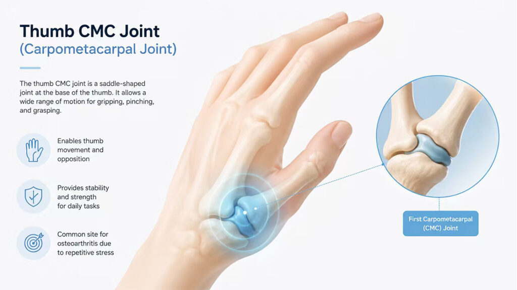
2.1 Why the Thumb Base Bears the Highest Load
The thumb CMC joint sits at the base of the thumb where it meets the wrist. This saddle-shaped joint provides the remarkable range of motion that allows the human hand to oppose, pinch, and grip. However, this same mobility comes with a trade-off: relatively less inherent stability compared to more constrained joints. During manual techniques requiring sustained pinching, gripping, or pressing, forces transmitted through the thumb CMC joint can reach levels many times greater than the external resistance applied. The joint capsule, ligaments, and supporting musculature work together to maintain alignment under load, but repetitive high-force demands accelerate cartilage wear.
2.2 Recognizing the Telltale Signs in Therapists
Physical therapists experiencing thumb CMC OA typically notice specific patterns of discomfort. Pain occurs at the base of the thumb during activities that require gripping, such as performing deep tissue work or holding a patient’s limb during mobilization. Patients often describe a deep ache that worsens with use and improves with rest. As the condition progresses, therapists may observe decreased grip strength and reduced ability to sustain manual contacts for extended periods. Pinching motions, particularly those requiring forceful opposition of the thumb and index finger, become increasingly uncomfortable. Some therapists also report a clicking or grinding sensation with thumb movement, indicating articular surface roughening.
2.3 The Functional Impact on Clinical Practice
Hand OA does not simply cause discomfort; it directly impairs a physical therapist‘s ability to perform essential clinical tasks. Sustaining manual contacts becomes more difficult as pain and fatigue set in earlier during treatment sessions. The reduced grip strength compromises the therapist’s ability to control joint mobilization forces precisely, potentially affecting treatment quality. Fine motor tasks such as palpating small anatomical structures, handling therapy instruments, or documenting notes on mobile devices also suffer. For therapists whose careers depend entirely on hand function, progressive OA represents a direct threat to professional longevity.
3. How Class IV Laser Therapy Modulates Tissue Response
Class IV laser therapy provides a non-invasive approach to supporting joint health and managing the inflammatory processes associated with osteoarthritis. Understanding the underlying mechanisms helps clarify how light-based treatment may benefit therapists dealing with occupational hand OA.
3.1 Photobiomodulation at the Cellular Level
At its core, Class IV laser therapy harnesses photobiomodulation. When specific wavelengths of light penetrate the skin and reach target tissues, photons interact with cellular components called chromophores. These chromophores, including cytochrome c oxidase found within the mitochondria, absorb light energy and trigger biochemical cascades. This activation increases cellular metabolism and supports the natural repair processes that maintain healthy joint tissues. The effect relies entirely on the body’s own physiological responses rather than introducing external substances or generating unnecessary tissue heating.
3.2 ATP Production and Cellular Energy
Mitochondria serve as the power plants of human cells, producing adenosine triphosphate (ATP)—the molecular fuel that drives everything from protein synthesis to ion transport. When Class IV laser light interacts with mitochondrial chromophores, the efficiency of ATP production increases. Cells with higher energy availability can perform their functions more effectively. For joint tissues, this translates into better maintenance of the extracellular matrix, more efficient removal of metabolic waste products, and enhanced capacity for responding to mechanical demands. Even small improvements in cellular energy balance can influence how joint structures tolerate daily loading.
3.3 The Inflammatory Response and Pain Signaling
Osteoarthritis involves both mechanical wear and inflammatory processes within the joint environment. Inflamed synovial tissues release various mediators that sensitize pain receptors and contribute to ongoing discomfort. Class IV laser therapy supports a reduction in inflammatory activity through multiple pathways. The light energy helps modulate the activity of pro-inflammatory mediators while encouraging the resolution phase of inflammation. Additionally, laser therapy influences nerve conduction by reducing the sensitivity of peripheral pain receptors, which contributes to decreased pain signal transmission without blocking all sensation. This dual effect on inflammation and neural signaling addresses both the tissue-level pathology and the symptom experience.
3.4 Deeper Penetration for Joint Structures
One of the distinguishing features of Class IV laser therapy lies in its penetration depth. Lower-power lasers may only reach superficial tissues, but Class IV devices deliver sufficient energy density to penetrate to joint capsules, deep tendons, and articular surfaces. For the thumb CMC joint, located beneath several layers of muscle, tendon, and soft tissue, adequate penetration is essential for therapeutic light to reach the target structures. The higher power output and appropriate wavelength selection allow clinicians to deliver meaningful energy doses to the joint itself rather than merely treating the overlying skin and subcutaneous tissues.
4. Integrating Laser Therapy Into Self-Care Protocols
Physical therapists who recognize the early signs of occupational hand OA can take proactive steps to support their long-term joint health. Incorporating Class IV laser therapy into a comprehensive self-care routine offers one avenue for managing tissue stress.
4.1 Recognizing When to Act
The optimal time to address hand discomfort is long before pain interferes with clinical work. Physical therapists should pay attention to morning stiffness lasting more than 15 minutes, achiness following busy treatment days, or reduced tolerance for manual techniques that previously caused no difficulty. These early signals represent opportunities for intervention. Waiting until pain forces modifications to treatment approaches often means the condition has already advanced. Therapists who treat their own early symptoms with the same attention they give their patients’ complaints typically achieve better long-term outcomes.
4.2 The Role of Recovery Periods After Clinical Sessions
After a day of delivering manual therapy, the therapist‘s hand joints have accumulated microdamage from hundreds of loading cycles. The post-clinic period provides a window for supporting tissue recovery before the next day’s demands begin. Applying Class IV laser therapy during this recovery window may help modulate the inflammatory response to daily mechanical loading. The goal is not to eliminate all inflammation—some inflammatory signaling is necessary for tissue repair—but rather to support an appropriate balance between breakdown and rebuilding. Regular laser sessions as part of an end-of-day recovery protocol may help maintain this balance over weeks and months of clinical practice.
4.3 Complementing Other Self-Management Strategies
Laser therapy works most effectively as one component of a broader self-care approach. Proper ergonomics during manual techniques reduces unnecessary joint loading. Taking brief rest breaks between patients allows tissues to recover partially before the next demand. Gentle range-of-motion exercises maintain joint mobility without provoking symptoms. Therapists should also consider how they position their hands during non-clinical activities, such as typing on keyboards or holding mobile devices, as these sustained positions add to cumulative tissue stress. When combined with Class IV laser therapy, these behavioral modifications create a comprehensive strategy for preserving hand function across a long career.
5. Practical Considerations for Occupational Joint Health
Maintaining hand health requires attention to daily habits, workplace strategies, and thoughtful planning for long-term career sustainability. Physical therapists who prioritize their own joint health serve both themselves and their patients.
5.1 Ergonomic Modifications During Patient Care
Small adjustments to manual therapy techniques can reduce joint loading without compromising treatment effectiveness. Therapists should consider using both hands symmetrically when possible rather than relying on a dominant thumb for all mobilizations. Supporting the patient‘s limb on a plinth or using body weight rather than isolated hand strength reduces the force transmitted through individual finger and thumb joints. When performing sustained pressure techniques, alternating hand positions or taking brief micro-breaks every few minutes prevents any single joint from experiencing prolonged loading. These modifications require conscious attention but become automatic with practice.
5.2 Building Recovery Into the Workday
The physical demands of clinical practice do not end when the last patient leaves. Documentation, equipment handling, and administrative tasks continue to load the hands throughout the workday. Therapists should structure their schedules to include brief recovery periods. Standing and stretching between patients, alternating between manual and non-manual tasks, and using voice recognition or dictation tools for documentation reduce the total time hands spend in active gripping or sustained postures. Even five minutes of rest every hour significantly reduces cumulative daily loading.
5.3 Long-Term Career Planning
Physical therapists planning for decades of clinical practice should consider how their hand health will evolve over time. The demands placed on hand joints at age 30 differ substantially from those at age 50, though many therapists continue using the same techniques throughout their careers. Gradually shifting toward techniques that place less stress on the small joints, incorporating technology such as therapeutic lasers or shockwave devices that reduce manual demands, and developing expertise in patient education and exercise prescription allow therapists to remain clinically effective even as their hands age. Planning for this transition before symptoms develop ensures a sustainable career trajectory.
Frequently Asked Questions
Q1: How can I tell if my thumb pain is early osteoarthritis or just overuse soreness?
Early morning stiffness lasting beyond 15 minutes and pain at the base of the thumb during pinching activities suggest OA rather than simple muscle fatigue.
Q2: Can I continue performing manual therapy techniques with thumb CMC OA?
You can with modifications, but continuing the same techniques without changes often accelerates joint degeneration. Adjusting hand positions and reducing loading helps.
Q3: How does Class IV laser therapy differ from low-level laser for hand OA?
Class IV lasers deliver higher power output, allowing deeper penetration to reach the thumb CMC joint and deliver meaningful energy doses in shorter treatment times.
Q4: How often should I use laser therapy for maintaining hand joint health?
Many therapists incorporate sessions after busy clinical days or several times weekly, but individual frequency depends on symptom severity and clinical demands.
Q5: Does Class IV laser therapy help with the cartilage changes of OA?
The therapy supports the cellular environment within the joint, including improving mitochondrial function and modulating inflammation, which benefits overall joint health.
Q6: Will laser therapy eliminate my hand pain completely?
The goal is supporting joint health and managing symptoms, not eliminating all sensation. Many therapists report improved tolerance for manual work with regular use.
Conclusion
Physical therapists face a genuine occupational risk of developing hand osteoarthritis, particularly at the thumb carpometacarpal joint, due to the repetitive, high-force demands of manual therapy practice. Understanding the mechanisms of articular degeneration and recognizing early warning signs allows therapists to take proactive steps before minor discomfort evolves into career-limiting impairment. Class IV laser therapy offers a science-based approach to supporting joint health through photobiomodulation, improved cellular metabolism, and modulation of inflammatory processes within the affected tissues. By combining appropriate ergonomic modifications, structured recovery periods, and thoughtful long-term planning, physical therapists can protect the hands that heal others while maintaining clinical effectiveness throughout their careers.
References
Nazari, H., Hosseini Mahjoob, H., Tapak, L., & Mortazavi, S. S. (2017). Prevalence of Work-related Musculoskeletal Disorders and Injuries in Occupational and Physical Therapists and Its Comparison.
https://irj.uswr.ac.ir/article-1-598-en.html
Cantero-Téllez, R., Kristin, V., Villafañe, J. H., & Medina Porqueres, I. Does laser therapy improve pain or pinch strength for thumb carpometacarpal joint osteoarthritis as an isolated treatment? A randomized controlled trial.
https://core.ac.uk/download/214833110.pdf
Medina-Porqueres, I., & Cantero-Tellez, R. (2017). Class IV laser therapy for trapeziometacarpal joint osteoarthritis: Study protocol for a randomized placebo-controlled trial.
Smart Laser Therapy. Laser Therapy in Physiotherapy.
https://smartlasertherapy.com/laser-therapy-in-physiotherapy
Medray Laser. What Is Class 4 Laser Therapy.